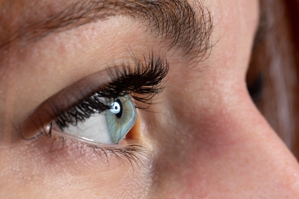
The retina is a delicate, light-sensitive layer of tissue lining the back of your eye. It plays a crucial role in your vision by converting light into electrical signals that the brain can interpret. When the retina becomes detached from the underlying tissue, it can lead to vision loss and even blindness if not treated promptly. Understanding the signs and symptoms of retinal detachment is essential for maintaining your eye health and preserving your sight.
What is Retinal Detachment?
Retinal detachment occurs when the retina separates from the back of the eye, much like wallpaper peeling off a wall. This separation can happen due to a variety of factors, including injury, age-related changes, or underlying eye conditions. When the retina becomes detached, it no longer receives the necessary oxygen and nutrients, leading to vision impairment and potential permanent vision loss if left untreated.
Common Causes of Retinal Detachment
Aging: As you grow older, the vitreous (the gel-like substance that fills the inside of the eye) can shrink and pull away from the retina, causing it to tear or detach.
Eye Injury: Trauma to the eye, such as a blow or puncture, can cause the retina to tear or become detached.
Nearsightedness (Myopia): People with high degrees of nearsightedness have a greater risk of experiencing retinal detachment due to the elongated shape of the eye.
Previous Eye Surgery: Certain eye surgeries, such as cataract removal, can increase the risk of retinal detachment.
Diabetes: Diabetic eye disease can weaken the blood vessels in the retina, making it more susceptible to detachment.
Recognizing the Symptoms of Retinal Detachment
Prompt recognition of the signs of retinal detachment is crucial, as early treatment can often prevent permanent vision loss. Some of the most common symptoms include:
Sudden Increase in Floaters and Flashes: You may notice an increase in the number of floating specks, cobwebs, or other shapes in your field of vision, accompanied by flashes of light.
Sudden Onset of Blurred or Distorted Vision: As the retina becomes detached, you may experience a sudden, noticeable change in your vision, such as blurriness, distortion, or a dark shadow or curtain in your peripheral vision.
Sudden Loss of Side (Peripheral) Vision: Retinal detachment often starts in the peripheral vision, causing a gradual loss of side vision that can eventually lead to tunnel vision.
Not all of these symptoms may be present, and the severity can vary. If you experience any of these signs, it is crucial to seek immediate medical attention from an optometrist.
The Importance of Regular Retinal Imaging and Dilation
Regular eye exams and retinal imaging are essential for the early detection and prevention of retinal detachment. Your optometrist can use specialized imaging techniques, such as dilated eye exams and Optomap retinal imaging, to closely examine the health of your retina and identify any potential issues before they progress.
Dilated eye exams allow your optometrist to get a better view of the retina and detect any abnormalities or changes. This examination is particularly important for individuals with risk factors for retinal detachment, such as high myopia, a history of eye injury, or a family history of the condition.
Optomap Retinal Imaging: A Breakthrough in Early Detection
Optomap retinal imaging is a revolutionary technology that captures a high-resolution, panoramic image of the retina in a matter of seconds. This advanced imaging technique allows your doctor to thoroughly examine the entire retina, including the peripheral areas, without the need for dilation.
The Optomap system provides several key benefits for the early detection of retinal detachment. The Optomap captures approximately 82% of the retina in a single image, allowing your eye care provider to thoroughly assess its overall health. The high-resolution Optomap images can help your doctor identify even the earliest signs of retinal abnormalities or changes, enabling prompt intervention and treatment if necessary.
By incorporating Optomap retinal imaging into your regular eye exams, your optometrist can better monitor the health of your retina and catch any potential issues before they lead to vision-threatening complications.
Prioritizing Your Eye Health at Vision Source Grove Heights
Retinal detachment is a serious condition that requires prompt medical attention to prevent permanent vision loss. By understanding the common causes, recognizing the early warning signs, and prioritizing regular retinal imaging and eye exams, you can take an active role in safeguarding your eye health.
If you have any concerns about the health of your retina or are due for a comprehensive eye exam, schedule a comprehensive eye exam with Vision Source Grove Heights. Our state-of-the-art Optomap technology and personalized care can help ensure the long-term health and function of your eyes. Visit our office in Houston, Texas, or call (346) 782-0288 to book an appointment today.


